
Secondary PPH, which is less common than primary PPH, is defined as any major bleeding occurring 24 hours to 6-12 weeks after delivery.3-5 For healthcare providers (HCPs) working in outpatient settings who see women who have recently given birth, the focus is on prompt recognition, diagnosis, and management of secondary PPH in order to improve maternal outcomes.
Causes of secondary postpartum hemorrhage
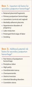
Common causes of secondary PPH include retained placental or fetal tissue, infection, and subinvolution of the placental site (delayed or inadequate physiologic closure and sloughing of the superficial modified spiral arteries at the placental attachment site).5,6 Less common causes of secondary PPH include bleeding disorders, uterine artery pseudoaneurysm, arteriovenous malformations, choriocarcinoma, cervical carcinoma, adenomyosis, diverticulum of the uterus, heavy menses, hypoestrogenism, and cesarean incision site dehiscence.5
Assessment
Initial assessment of secondary PPH focuses on the woman’s hemodynamic stability. If she is deemed unstable—for example, she has S/S of hypovolemia and substantial ongoing bleeding—immediate transport to an emergency care facility is required. Oxygen should be administered and two large-bore intravenous (IV) lines should be established as soon as possible for fluid replacement with a crystalloid solution until blood is available.4,7 If the patient is deemed stable, the HCP should obtain a thorough history and conduct a physical examination to assess potential causes of bleeding. In addition, the HCP should continue to monitor the patient’s hemodynamic status.
The history focuses on three primary areas: severity of bleeding, any previous interventions, and potential predisposing factors. The
 HCP needs to assess the severity of the bleeding by asking the patient about the amount of immediate post-delivery bleeding, the pattern of bleeding since delivery, and the current amount of bleeding. Bleeding that soaks through at least one pad per hour and/or the passing of blood clots the size of an egg or larger represents excessive bleeding.4 Objective quantification of blood loss, albeit superior to observed or reported quantification, may be difficult to obtain in secondary PPH. Objective quantification of blood loss involves an equation that measures the dry gram weight of an item and subtracts it from the wet gram weight of an item; the result of that equation is equivalent to the milliliters of blood lost.8
The HCP should perform a complete physical examination, looking especially for S/S of hypovolemia, including tachycardia, hypotension, tachypnea, weak peripheral pulses, delayed capillary refill, pallor, skin mottling, and altered mental status, as well as S/S of coagulopathy (e.g., petechiae, bruises, mucosal bleeding).4 The HCP should palpate the abdomen for tenderness, distention, uterine enlargement (may indicate atony), and bladder overdistension.4 During the perineal and speculum exam, the HCP should carefully evaluate the current status of bleeding, as well as the presence and extent of lacerations, hematomas, and malodorous lochia. A bimanual examination should be performed to assess for uterine bogginess, enlargement, or tenderness.4 The HCP should be aware that, on occasion, the fundus can be firm and contracted down, but atonic, in the lower uterine segment.3 In this scenario, the HCP can remove clots and perform bimanual compression to reduce blood loss until uterotonic agents are available and begin to work.3
Pelvic ultrasound and laboratory tests may assist in determining the cause of the bleeding. Ultrasound may reveal retained placental fragments. However, the HCP needs to keep in mind that, on ultrasound, the uterus of a patient with secondary PPH may appear similar to that of a normal postpartum uterus.5 Laboratory tests should include a complete blood cell count, pro-thrombin time, activated partial thromboplastin time, fibrinogen level, and quantitative human chorionic gonadotropin.5
Management
When secondary PPH is suspected, prompt consultation with an obstetrician/gynecologist is necessary in the event that advanced interventions might be needed. While management of the patient is being discussed, the HCP should make arrangements for hospital admission so that these advanced interventions can be initiated.
Patients with secondary PPH may not experience typical signs of anemia: Hemoglobin and hematocrit values may not accurately reflect the amount of blood loss, and vital signs may not change until substantial blood loss occurs.5 If suspected blood loss is 1,500 mL or more and vital signs are unstable, immediate preparation for blood transfusion should be made.5 Pharmacologic management o f subinvolution of the placental site involves the use of uterotonic agents: methylergonovine 0.2 mg intramuscularly (IM) every 2-4 hours for up to three doses, carboprost tromethamine 250 mcg IM every 15 minutes for up to eight doses, and/or oxytocin by intravenous infusion.5 If pharmacologic management fails to control bleeding, the patient may require surgical intervention (dilation and curettage [D&C], suction curettage). Some patients may not respond to pharmacotherapy or curettage, and a hysterectomy may be necessary.5
Of note, hemorrhage occurring 2-5 days postpartum may indicate the presence of von Willebrand disease (VWD).4 During this postpartum period, Factor VIII levels that had increased in pregnancy begin to decrease.4 Patients with VWD may be at risk for late PPH until 4 months postpartum.4 HCPs who suspect the presence of VWD or other coagulopathy in a given patient should refer her to a hematology specialist for further evaluation.
Postpartum discharge education
Prior to discharge, education of new mothers and the persons who support them regarding S/S that could indicate PPH—or any other postpartum complication—is an important measure in preventing severe morbidity and mortality in the postpartum period. A simple mnemonic, POST-BIRTH, which includes the S/S of PPH and other potentially life-threatening complications, was developed by the Association of Women’s Health, Obstetric and Neonatal Nurses (AWHONN) for providers and birthing facilities to distribute to postpartum mothers.11 POST-BIRTH stands for:
• Pain in chest
• Obstructed breathing or shortness of breath
• Seizures
• Thoughts of hurting yourself or your baby
• Bleeding, soaking through one pad/hour, or blood clots the size of an egg or bigger
• Incision that is not healing
• Red or swollen leg that is painful or warm to touch
• Temperature of 100.4°F or higher
• Headache that does not get better, even after taking medicine, or bad headache with vision changes
This list of S/S provides guidance for patients on when to call 911 (POST) and when to call the provider (BIRTH).10,11 Patient and family awareness is vital in the early recognition of and intervention for PPH and the prevention of subsequent severe morbidity and mortality.
Conclusion
Postpartum hemorrhage remains a major cause of morbidity and mortality in the U.S. Whereas primary PPH accounts for the vast majority of cases, HCPs in primary care are more likely to encounter patients with secondary PPH. Secondary PPH is defined as any significant uterine bleeding that occurs 24 hours to 12 weeks post-delivery. HCPs must be able to recognize secondary PPH and be prepared to manage these patients. First, HCPs who suspect secondary PPH should consult with an obstetrician/gynecologist in anticipation of advanced interventions. A detailed history focusing on blood loss, previous interventions, and potential causes should be obtained. The physical exam should focus on signs of hypovolemia, followed by an abdominal and pelvic exam. Management depends on the cause and severity of blood loss. Treatment may range from administration of uterotonic agents to surgical intervention. In an effort to prevent serious complications, patients should be educated about PPH and know when to call 911 or their HCP.
2. World Health Organization. WHO Recommendations for the Prevention and Treatment of Postpartum Haemorrhage. 2012. apps.who.int/iris/bitstream/10665/75411/1/9789241548502_eng.pdf
3. American College of Obstetricians and Gynecologists. Practice Bulletin #183. Postpartum Hemorrhage. Washington DC: ACOG; 2017.
4. Chye TT, Tagore S. Puerperal problems. In: Janes D, Steerm PJ, Weiner CP, et al, eds. High-Risk Pregnancy: Management Options. 5th ed. New York, NY: Cambridge University Press; 2017:1966-1985.
5. Belfort MA. Secondary (late) post- partum hemorrhage. UpToDate. May 21, 2018. uptodate.com/contents/secondary-late-postpartum-hemorrhage/print
6. Weydert JA, Benda JA. Subin- volution of the placental site as an anatomic cause of post-partum uterine bleeding: a review. Arch Pathol Lab Med. 2006;130(10):1538-1542.
7. Government of Western Australia Department of Health. Second- ary Postpartum Haemorrhage. 2016. kemh.health.wa.gov.au/~/media/Files/Hospitals/WNHS/For%20health%20professionals/Clinical%20guidelines/OG/WNHS.OG.PostnatalComplicationPPHSecondary.pdf
8. Association of Women’s Health, Obstetric and Neonatal Nurses. Quantification of blood loss: AWHONN practice brief number 1. JOGNN. 2014;44(1):158-160.
9. Karsnitz DB. Postpartum compli- cations. In: King TL, Brucker MC, Kriebs JM, et al, eds. Varney’s Midwifery. 5th ed. Burlington, MA: Jones & Bartlett Learning; 2015:1143-1156.
10. Supplee PD, Kleppel L, Santa-Do- nato A, Bringham D. Improving postpartum education about warning signs of maternal morbidity and mortality. Nurs Womens Health. 2016-2017;20(6):552-567.
11. Association of Women’s Health, Obstetric and Neonatal Nurses website. POST-BIRTH Education Program. 2018. awhonn.org/page/POSTBIRTH